A labelled brain diagram can feel manageable until the labels disappear. Then “temporal,” “parietal” and “occipital” become a list of words with no reliable location or meaning. Memorising the same picture again may strengthen recognition without fixing the missing connections.
Build each revision item around three prompts instead: Where is the region? What is one important association? What would be misleading to claim about it? The third question matters because the brain does not operate as a collection of isolated departments, each responsible for exactly one behaviour.
Make a small map before adding detail
Start with the four cerebral lobes. The frontal lobe lies towards the front, the parietal lobe behind it, the temporal lobe towards the side, and the occipital lobe towards the back. As introductory anchors, connect frontal areas with motor control and aspects of executive processing, parietal areas with somatosensory processing, temporal areas with auditory processing, and occipital areas with vision. These are starting associations, not complete job descriptions. OpenStax, Psychology 2e, Brain and Spinal Cord
Use a diagram approved for your course and check its orientation. A side view, a top view and a section through the brain do not show the same structures equally well. Write “left lateral view” or the appropriate orientation beside your practice image before labelling it.
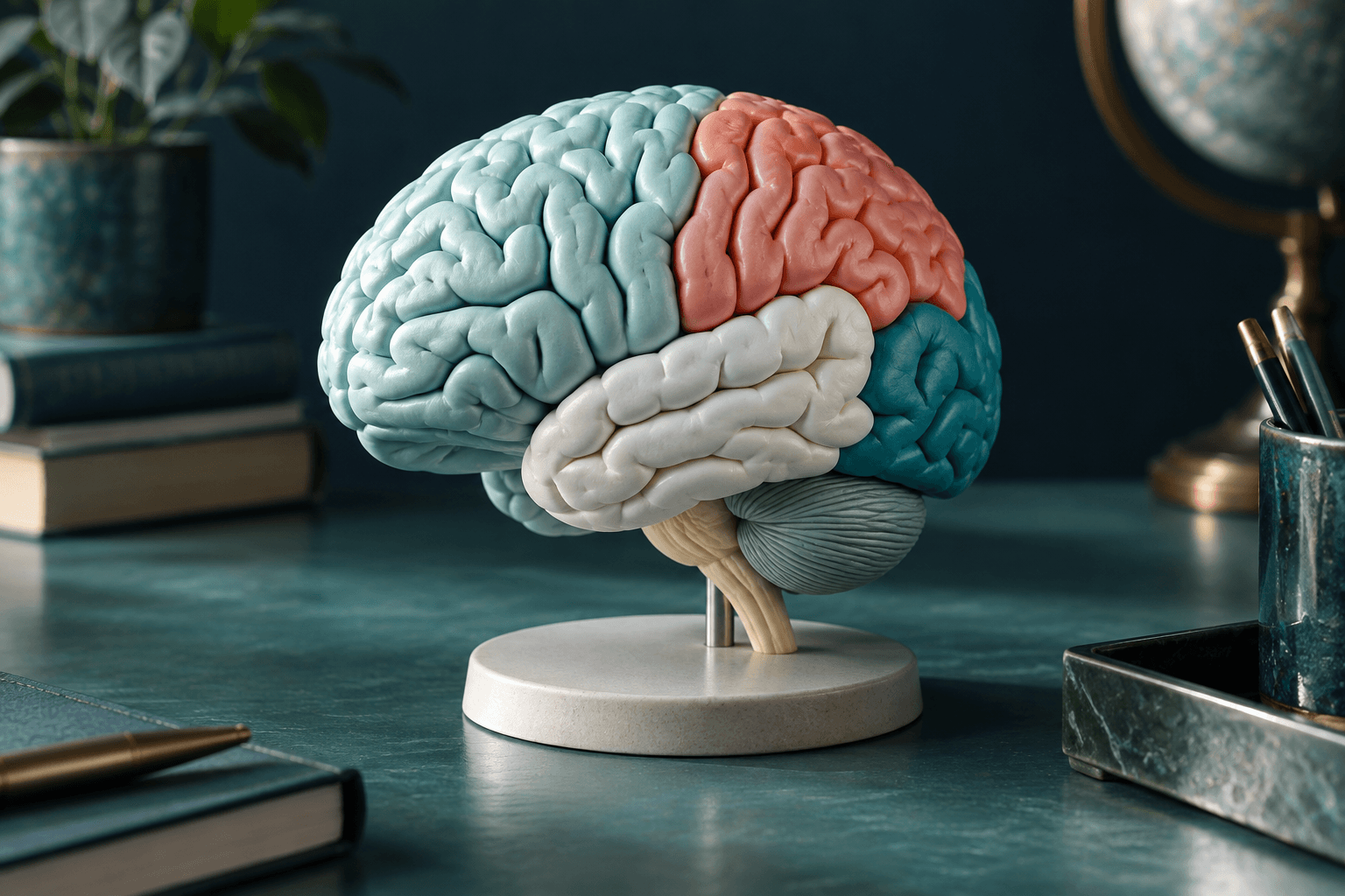
Do not use an AI-generated decorative brain model as your anatomical answer key. A visually plausible fold is not evidence that the boundary or label is correct. Keep decorative imagery separate from the diagram you use to learn anatomy.
Add structures without assigning them a single personality
The hippocampus is important for learning and memory, and the hypothalamus is involved in homeostatic regulation. Those associations help organise introductory study, but statements such as “all memory is stored in the hippocampus” are too broad. Different tasks depend on interacting systems. OpenStax, Brain and Spinal Cord
BrainFacts describes neural circuits as connected processing systems, including recurrent connections that provide feedback. This is a useful counterweight to the one-region–one-function habit. A region can make a specialised contribution while still depending on information from elsewhere. Society for Neuroscience, 2022
On a revision sheet, leave space for “works with” beside “associated with.” You do not need to draw every connection. The blank itself reminds you that a functional label is not a claim of independence.
Worked example: improve a weak flashcard
Consider a card that says “Occipital lobe = eyes.” It gives you a memorable word pair but confuses a brain region with a sensory organ. It also says nothing about where the lobe is.
Replace it with three prompts: “Locate the occipital lobe on this lateral view”; “Name a major processing association”; and “Why is it inaccurate to say this region works alone whenever someone reads?”
The answers should identify the posterior location, visual processing, and the need for other processes in the wider task. Reading is not merely detecting marks: the exercise may also involve language and meaning. You do not need to invent a single exact pathway to recognise that the task crosses your simple revision categories.
Now make a reverse card. Give the function prompt first and ask which region is a useful introductory association. Practising both directions helps reveal whether you learned the relationship or only the order of a familiar list.
Compare neighbouring ideas
Create a two-column comparison when names repeatedly collide. For example, place parietal and temporal on the same page. Add location, one association and a small sketch. Then cover the names and explain the difference aloud.
Next, remove the helpful pairing and mix these prompts with other regions. A card that is easy when its only alternative is visible may be harder in a larger set. Record that difference rather than treating every correct answer as equivalent evidence of readiness.
This approach is an exercise design, not a claim that one exact card format is scientifically optimal. Research on retrieval practice supports testing memory rather than relying entirely on repeated exposure, but performance depends on the task and conditions. Roediger and Karpicke, 2006
Keep case questions within their scope
An introductory examination may give a simplified description and ask for a likely anatomical association. Answer the question at that level, while recognising that real clinical interpretation requires much more information. Do not use a study quiz to diagnose yourself or another person.
A useful written-answer pattern is: identify the relevant association, point to the detail in the question, and state the limitation if the prompt asks for evaluation. This avoids two extremes: naming a region with no explanation, or adding a confident diagnosis that the evidence cannot support.
A short self-test
1. Why is “frontal lobe = personality” an incomplete revision card?
Answer: It is broad, does not specify a process or location, and suggests an isolated responsibility. Replace it with a more precise association required by your syllabus.
2. You can label a side-view diagram but fail on a section. What should you practise?
Answer: Identify the orientation and use an approved view that actually shows the target structure. Do not assume the same visual landmarks transfer unchanged.
3. Does identifying an association prove that one region alone performs the whole task?
Answer: No. Specialisation and interaction can coexist. A simple map is an entry point, not a complete network model.
4. Why keep a limitation on a flashcard?
Answer: It prevents a useful shortcut from becoming an absolute claim. Knowing what a label does not establish is part of understanding it.
Choose four regions and build location, function and limitation prompts for each. For a future study option, get notified about Psych Explained’s upcoming Neuroscience course. It is not available yet; use your existing course resources while waiting for launch information.
Sources and further reading
- Spielman, R. M., Jenkins, W. J., and Lovett, M. D. (2020). Psychology 2e, section 3.4, The Brain and Spinal Cord. OpenStax.
- Society for Neuroscience (2022). Signaling Within Neural Circuits. BrainFacts.
- Roediger, H. L., and Karpicke, J. D. (2006). Test-enhanced learning. DOI.